Introduction
Blood transfusion is an accepted life saving therapeutic procedure in obstetrics, all specialities of medicine and few chronic or immune disorders of dermatology like chronic vascular disease and bullous dermatosis.1 Adverse dermatological reactions in the form of generalized purpura may occur mainly in women within 2-7 days post blood transfusion.2 Acute Graft versus host disease (GVHD) remains most dangerous life threatening adverse dermatological reaction following blood transfusion.3 Usually GVHD occurs following bone marrow/s olid organ transplantation but post blood transfusion, Acute GVHD may also occur. Chronic GVHD post Transfusion Associated GVHD is rare, an undiagnosed life threatening disease. However, rarely it may report as chronic GVHD.4 Post transfusion GVHD in a pregnant women is a rarity a chance occurrence. We report a unique adverse skin manifestation, painful extensive bullous and ulcerative lesions over lower abdomen and vulva with massive vulval oedema, high grade fever, thrombocytopenia, liver dysfunction and haemoglobinuria following acute blood transfusion reaction in an unbooked 28year G3 P2 L2 pre gnant woman with severe anaemia, first of its kind in my 31 years of clinical experience as consultant obstetrician.
Case Report
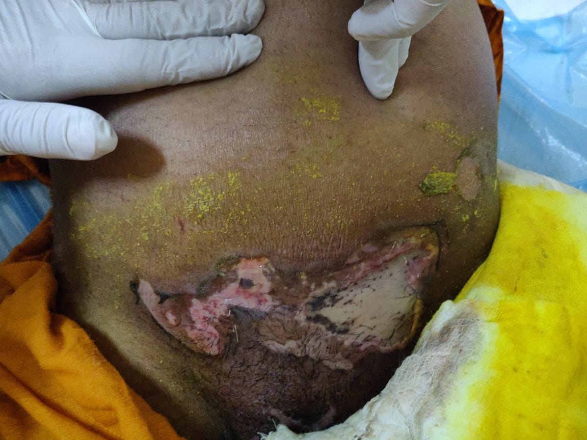
28Year, G3 P2 L2 unbooked with 29 weeks 2 days period of gestation was referred to the Emergency department of MMIMSR hospital, Mullana, Ambala, Haryana on 14/11/2018 with chief complaints of amenorrhoea seven months, fever since 2 days with severe anaemia and painful bullous eruptions over lower abdomen and vulva with massive vulval edema. Fever was sudden in onset, high grade with chills and rigors. There were no bowel or bladder complaints associated with fever. On general physical examination, pallor was +++, Blood pressure - 90/60mmHg, Pulse – 120 beats per minute, Respiratory rate was 28 per minute, was febrile with temp of 100.2 0 F. Patient was dyspnoeic and SPO 2 =90% on room air. On local examination, uterus was 30 weeks size of pregnancy, longitudinal lie and FHS was 160bpm and regular. There were extensive bullous and ulcerative skin lesions extending from one iliac fossa to other with deep excavating margins with serous ooze (Figure 1). There was massive vulval edema with ulcerative lesions (Figure 2). With rupture of bullae, skin lesions appeared dusky erythematous with necrotic plaques and areas of sloughing and ulceration. Skin lesions were tender to touch and painful. A week prior to reporting in our emergency, p atient was admitted to primary care private hospital where two units of blood were transfused in view of severe anemia. Following blood transfusion, patient went into shock with blood pressure of 70/40 mmHg and tachycardia. Shock was managed with inotropes (Noradrenaline nd dopamine) and oxygen inhalation as per the referral slip. Patient also developed high-grade fever with rigors and chills along with painful extensive bullous lesions over lower abdomen and vulva. There was no history of any drug allergy, insect bite, trauma or burns over lower abdomen/external genitalia. Haemoglobin was 7.0gm%, Bleeding time - 2.15 min, Clotting time- 6.30 minutes, TLC - 16,100/cumm with DLC -80,16,2,2. There was thrombocytopenia with platelet count 54,000/ uL. Peripheral blood film for Malarial Parasite, dengue serology and widal test were negative. Liver F unction Tests were raised. Total Bilirubin :3.7mg%, direct bilirubin: 3.0mg%, indirect:0.7mg%, SGPT:200IU/L, SGOT:104IU/L, Serum Alkaline Phosphatase : 309IU/L,S.LDH:448IU/L. Also, there was evidence of haemoglobinuria on urine microscopic examination pointing towards adverse blood reaction. HBsAg, HCV, HIV and VDRL were negative. Patient was given symptomatic treatment in collaboration with dermatology and medicine department. Patient was put on tablet prednisolone 40mg OD, intravenous fluids, intermittent oxygen and triple intravenous antibiotics (Augmentin, metronidazole and gentamycin) along with topical steroids and emoliont creams. There was remarkable improvement with marked regression of skin lesions patient was discharged in satisfactory condition after two weeks.
Figure 1
Image showing extensive bullous and ulcerative skin lesions extending from one iliac fossa to other.

Discussion
Adverse dermatological lesions following blood transfusion reaction in obstetrics are rarely seen. Generalised purpuric rash over body within two to seven days after blood transfusion has been reported.2 Graft versus host disease (GVHD) after blood transfusion is a rare dangerous, life threatening entity reported in literature. It can occur in women deficient in cell mediated immunity.3 GVHD usually occurs 2-30 days after blood transfusion. It presents as fever accompanied with multiorgan systemic manifestations with widespread exanthematous over the body with marked leucopenia. Our patient also had fever with extensive painful skin lesions over lower abdomen and vulva. LFTs were raised along with thrombocytopenia and haemoglobinuria. The mechanism responsible for adverse dermatological manifestation may be complex due to autoimmune reaction, or adoptive transfer of immunity to the recipient, Koebner’s phenomenon or microchimerism.1 Purpuric phototherapy induced eruption following blood transfusion has been reported in neonates.5 Release of porphyrins by haemolysis of erythrocyte precursors following blood transfusion may lead to raised levels of coproporphyrin and protoporphyrins in neonates.
Extensive purpuric skin lesions along with hypertension and convulsions has also been reported in literature following blood transfusion.6 Adverse periocular skin reaction following blood transfusion has been reported by Margo CE in 1999.7
In patients with cold agglutinin disease cutaneous necrosis at the site of blood transfusion has also been reported,8 and skin lesions in our patient also had necrotic plaques with areas of sloughing and ulceration.
Martin L et al in 2001 reported vitiligo and Sjogren’s syndrome in a woman after blood transfusion due to microchimerism.9 Fever with extensive adverse dermatological lesions, thrombocytopenia, haemoglobinuria and raised LFTs following blood transfusion were observed in our pregnant patient with severe anaemia.
Conclusion
Blood transfusion should be utilized only as a life saving procedure at secondary and tertiary care hospitals only. High index of suspicion and vigilant observation for any adverse dermatological reaction must be a consideration. High dependency unit ready to tackle with emergency crisis should be available in the same set up while transfusing blood and blood products.


