- Visibility 39 Views
- Downloads 4 Downloads
- DOI 10.18231/j.ijogr.2022.024
-
CrossMark
- Citation
Acute prolapse of giant submucosal fibroid polyp mimicking uterine inversion- A rare case report
Introduction
Leiomyoma is the commonest of all uterine and pelvic tumours, with an incidence of almost 20% in women of reproductive age group.[1] Fibroids can develop anywhere within the muscular wall of the uterus including submucosal, intramural, or subserosal sites. Inferquently, pedunculated submucosal fibroid may prolapsed into to vagnia through dilated cervical canal through uterine contractions. This can frequently present with heaviness in vagina, masss in the vagina or sensation of something coming out of the vagina or offensive excessive vaginal discharge. At times it is big enough to distend the vagina or even comes out of the introitus confusing the diagnosis of uterine inversion.[2] Usually polyps measure less than 2cm but if they are greater than 4cm they are considered as giant polyps.[3] Huge fibroid-polyp mimicking uterine inversion can cause diagnostic dilemma as it is rarely encountered in gynecological practice. Here we are reporting a case of prolapsed giant fibroidal polyp outside the introitus resembling uterine inversion presenting at the Department of OBG BLDE (DU) Shri BM Patil Medical College, Vijayapura.
Case Report
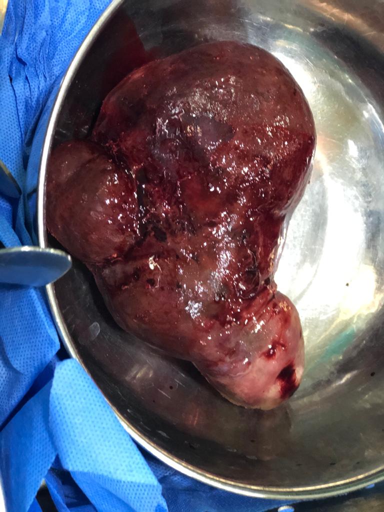
A 35-year old woman Para 2 Living 2 presented history of sudden descent of mass per vagina along with sudden gush of bleeding when she carried heavy weight as a part of her household activity. On admission, she had pallor with feeble pulse of110/min and blood pressure 130/80 mmHg and. Abdomen was soft and non tender. Local examination revealed a foul smelling irregular broad mass was lying outside the vulva([Figure 1]). The mass was bleeding. On bimanual examination, a pedicle of the mass was palpated arising from the uterine body. Cervical os could be delineated. Resuscitation was initiated. Her hemoglobin (Hb) was 7.2 g/dl. Ultrasonography revealed a normal sized and shaped uterus. The patient was shifted to the Operation Theater for evaluation and treatment. Under Subarachanoid block the patient was put in Lithotomy position.
Evaluation under anesthesia revealed that uterus was inside the pelvis. It was normal and size and shape. From the uterine body a long pedicle was attached to the growth. At the introitus a large irregular mass measuring 15cm x 13 cm × 10cm with areas of hemorrhage and necrosis was seen([Figure 2]). It was firm in consistency. The mass bled on touch. A diagnosis of pedunculated uterine fibroid polyp.was made. It was doubly clamped, cut using monopolar cautery and ligated with Polyglactin 910 No.1. Foley’s bulb was kept in situ inside the uterus to prevent hemorrhage and to achieve pressure hemostasis. The mass was sent for histopathological evaluation. Post operatively anaemia was corrected and antibiotics were given.
Histopathology Report showed features suggestive of leiomyoma with secondary changes.


Discussion
Uterine fibroid is the most frequent benign tumor of the female genital tract. Submucosal fibroids, which can be sessile or pedunculated, give more symptoms like heavy menstrual bleeding, intermenstrual bleeding, hydrorrhea, pain may be due to the stalk of the pedunculated type is twisted.[4] They impair the pelvic anatomy by pulling down the uterus in relation with the bladder and the rectum. And also the myoma becomes necrotic and infected cause of inadequate blood circulation through a long pedicle. If the pedicle of submucosal leiomyoma twists, infarction results, occasionally red, hemorrhagic, gangrenous degeneration occurs.[5] Till now in literature very few cases of giant cervical fibroid polyp are mentioned and are rare. Our case report is the only one that describes about acute prolapse of giant submucosal fibroid polyp.
Examination under anaesthesia, ultrasound and histopathology helped us in the diagnosis. A careful clinical and radiological examination prevents the unnecessary hysterectomies, as in our case polypectomy alone helped the patient. Written and informed consent was taken from the patient for publication of this case report and accompaying images.
Source of Funding
None.
Conflict of Interest
The authors declare no conflict of interest.
References
- N Bhatla. Tumous of the corpus uteri. Jeff Coates principles of Gynaecolgy 2001. [Google Scholar]
- JS Drinville, S Memarzadeh, AH De Cherney, L Nathan. Benign disorders of the uterine corpus. Current obstetrics and gynaecologic diagnosis and treatment 2007. [Google Scholar]
- S Das. Giant fibroid polyp mimicking uterine inversion. J Sci Soc 2020. [Google Scholar]
- S Sulaiman, A Khaund, N Mcmillan, J Moss, M A Lumsden. Uterine fibroids-do size and location determine menstrual blood loss?. Eur J Obstet Gynecol Reprod Biol 2004. [Google Scholar]
- I Bezircioglu, DK Sakarya, MH Yetimalar, E Kayhan, A Yildiz. A Huge Vaginal Prolapsed Pedunculated Uterine Leiomyoma: A Case Report. J Clin Stud Med Case Rep 2015. [Google Scholar] [Crossref]
