- Visibility 34 Views
- Downloads 3 Downloads
- DOI 10.18231/j.ijogr.2022.026
-
CrossMark
- Citation
Uterine rupture: A rare complication of repeated cervical cerclage
Introduction
Uterine rupture is a life threatening emergency which demands mindful obstetric practice or else it can lead to catastrophic consequences. The majority of uterine ruptures occur due to previous surgery on the uterus involving the myometrium including hysterotomy, cesarean section, myomectomy and adenomyomectomy. Rupture in an unscarred uterus is rare and completely unpredictable, the reported risk of rupture being approximately 22-74/10000 in previously scarred uteri and 0.5-2/10000 in unscarred uteri.[1] We report this case to highlight the rare event of rupture in an unscarred uterus and emphasize that maternal and fetal risk in this situation is directly related to prompt recognition and immediate surgical intervention.
Case History
Our patient, thirty-six years old gravida 3 para 1 abortion 1, presented in early labor at 36+5 weeks of gestation. Her medical and surgical history was not significant. During the first pregnancy, a spontaneous twin conception, an emergency cervical cerclage was performed at 20 weeks, but was subsequently removed due to preterm premature rupture of membranes, followed by second trimester abortion at 22 weeks. During the second pregnancy, cervical cerclage was done at 16 weeks of gestation due to cervical incompetence. The cerclage was removed at 36 completed weeks, and she had a spontaneous vaginal delivery a week later.


In this pregnancy, the antenatal period was uneventful. Based on her past obstetric history, cervical cerclage was performed by McDonald’s technique at 14 weeks gestation with a plan to remove the cerclage at 36 completed weeks. But at 36+5 weeks, she presented in labor and was admitted to hospital with mild contractions. Her vitals were stable and an admission test documented a baseline fetal heart rate of 136-140 bpm, baseline variability of 8-10 bpm with 5 accelerations in 20 minutes and no decelerations.
The cerclage was removed and the cervix immediately dilated to 3 cms and was 60% effaced with the presenting part stationed at -2. The patient was monitored and labor was allowed to progress spontaneously. After 2 hours the membranes ruptured spontaneously and per vaginal examination revealed clear liquor with the cervix 4 cms dilated and 80% effaced, with the vertex at occipito transverse position and the station at -1. Within 30 minutes, the contractions picked up in intensity, duration and frequency. The patient was administered epidural analgesia for pain relief at request. Continuous monitoring of fetal heart rate and patient vitals was done and partogram was charted. After 3 hours, early deceleration of fetal heart rate to 86 bpm were noted with immediate pick up to 126 -132 bpm. On per vaginal examination the cervix was fully dilated, the vertex at occipito anterior position and the station +1. The early decelerations were attributed to head compression and patient was encouraged to bear down.
In less than 5 minutes, there was a prolonged deceleration upto 60 bpm with no pick up in spite of resuscitative measures which included left lateral position, intravenous fluid infusion and oxygen by mask at 4-6 litres flow.
The patient and her family were appraised of the emergency, and she was shifted to the operation theatre with a recorded consent for instrumental delivery or an emergent caesarean section as the situation demanded. There was persistent fetal deceleration to less than 60 bpm and a per vaginal examination in the operation theatre revealed a regression of station to above -1. With a high suspicion of uterine rupture, an immediate caesarean section was performed under epidural anesthesia. On opening the abdomen, hemoperitoneum of approximately 800 ml was found with active bleeding from the left side of the lower uterine segment. Baby was delivered and handed over to neonatologist. The baby cried after 45 seconds of bag mask ventilation with Apgar scores of 4 and 8 at 1 and 5 minutes respectively.
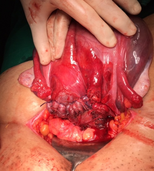
In view of tachycardia and hypotension, general anesthesia was administered and patient was resuscitated with a rapid infusion of intravenous crystalloids and colloids. The uterus was exteriorized and a complete tear of approximately 6 cms was seen on the lateral aspect of the lower segment, extending from the upper cervix to the left side of the uterus. A repair of the rent was undertaken with No. 1 polyglactin 910 using continuous locking along with a few interrupted sutures.
Intraoperatively, two units of packed cells were transfused along with intravenous 1gm tranexamic acid, intravenous 20 units oxytocin infusion and 250 mcg intramuscular 15 methyl PGF2 alpha to control bleeding and prevent post partum hemorrhage. There was persistent uterine atony for which B Lynch compression sutures were taken and hemostasis was confirmed. The final blood loss measured from the suction aspirate and soakage of mops and under sheets was estimated to be 1500 ml. Postoperatively, patient was monitored in high dependency unit for 24 hours. The postoperative recovery was uneventful and the patient was discharged with her baby on day 7.
Discussion
Though the incidence of rupture in unscarred uterus is rare, there are many cases reported in literature with varied etiology. Maternal connective tissue disorders are a known cause for rupture in unscarred uterus and patients with Ehler Danlos Type IV are at high risk due to the congenital weakness of the uterus.[2] Uterine anomalies are also potential risk factors and rupture of 4 cases of unscarred bicornuate uterus have been reported by Walsh et al. in their study of uterine rupture in primigravidas.[1] Other causes of rupture of unscarred uteri include prolonged labor, cephalopelvic disproportions, fetal macrosomia, postdated pregnancies and malpresentations.[3] Complications associated with obstetric interventions like internal podalic version, vaginal breech extraction and instrumental deliveries are the iatrogenic causes of rupture of the unscarred uterus.[4]
Combination induction and augmentation of labor had the highest risk of uterine rupture, followed by prostaglandin induction and then by oxytocin induction. Contributing factors include high parity, advanced maternal age and short interval between deliveries.[1]
Vernekar et al in their retrospective analysis of unscarred uterine rupture reported 10 cases of rupture at term. In their study, the rupture occurred due to mismanaged labor (30.8%), the use of oxytocin (23%), instrumental delivery (15.4%), obstructed labor (15.4%), induction by prostaglandin gel (7.7%) and placenta percreta (7.7%).[5] Kuba et al published a case report with prior uterine instrumentation being the cause of rupture in unscarred uterus.[6]
In our case, due to absence of any of the above factors, we believe repeated cervical cerclage to be the cause of rupture. Repeated cervical cerclage may cause fibrosis and scarring of the cervix. The stretching capacity of the cervix is compromised leading to subsequent uterine tear. The forces and pressures of active labor may then cause the rent to extend upwards into the lower uterine segment. In literature, we could find three studies where cervical cerclage was considered to be the cause of this life threatening entity. Ogawa et al have described a uterine rupture in a case of previous two cerclages following a cervical conization procedure.[7] Nashar et al have described a uterine rupture in a case of previous six cerclages.[2] Fox et al in their study of labor outcomes in 69 patients after Shirodkar cerclage reported two cases of uterine rupture of an unscarred uterus.[8] As in our case, the second stage of labor is reported to be the most common stage of uterine rupture by Aggarwal et al.[2]
Consequences of uterine rupture depend upon the time interval between the actual occurrence of the rupture, diagnosis and intervention.[9] Maternal deaths and perinatal deaths were 30.8% and 53.8% respectively as per Vernekar et al in their study.[5] Kuba et al have reported maternal and fetal mortality of 16% and 83% respectively in unscarred uterus which is higher than previously scarred uterus.[6] Gibbins et al, in a case control study, compared the outcomes of uterine rupture in 20 cases of unscarred uterus to 120 cases of scarred uterus. They reported that primary uterine rupture had greater maternal morbidity, greater mean blood loss, higher rate of blood and blood products transfusion and higher rates of obstetric hysterectomy as compared to cases of previously scarred uterus.[10] Early diagnosis and surgical intervention are the key to successful management of uterine rupture. The hallmark signs of uterine rupture are loss of fetal station along with fetal bradycardia, maternal tachycardia and hypotension. Other symptoms include abdominal pain and vaginal bleeding. In our patient, fetal bradycardia and loss of fetal station pointed towards the diagnosis of uterine rupture. We shifted the patient to the operation theatre because of the prolonged deceleration anticipating an obstetric complication. The management of suspected uterine rupture is laparotomy with cesarean delivery followed by correcting the cause and management of hemorrhage. Management decisions whether to repair the rent or opt for an obstetric hysterectomy have to be individualized by clinical condition, degree and location of tear, hemorrhage and surgical complications as also desire for future child bearing.[6]
Conclusion
This case is a demonstration of how an apparent low risk situation can turn critical within minutes in obstetrics. Heightened awareness, close supervision and optimum threshold for intervention would enable obstetric teams to achieve desired results while facing this or any other obstetric emergency. A high index of suspicion, prompt diagnosis, timely intervention and a multidisciplinary approach is what helped us salvage a critical condition and avert an adverse fetal and maternal outcome in our case.
Source of Funding
None.
Conflict of Interest
The authors declare no conflict of interest.
References
- A Bharti, P Singh, N Nalini. Posterior uterine wall rupture during labour in primigravida - A rare case report. Int J Contemp Med Res 2018. [Google Scholar]
- S Dubey, J Rani, M Satodiya. Unexpected Uterine Rupture: A Case Series and Review of Literature. J Clin Diagn Res 2018. [Google Scholar]
- S D Halassy, J Eastwood, J Prezzato. Uterine rupture in a gravid, unscarred uterus: A case report. Case Reports in Women’s. Health 2019. [Google Scholar]
- SM Yussof, CC Hsuan. An Unusual Presentation of Rupture in an Unscarred Uterus. Crit Care Obst Gyne 2016. [Google Scholar] [Crossref]
- M Vernekar, R Rajib. Unscarred Uterine Rupture: A Retrospective Analysis. J Obstet Gynecol India 2016. [Google Scholar]
- K Kuba, DN Matseoane-Peterssen, D Goffman. Rupture of an Unscarred Uterus in a Woman with Recurrent Prior Uterine Instrumentation. J Clin Obstet Gynecol Infertil 2018. [Google Scholar]
- M Ogawa, Y Konishi, M Obara, T Tanaka. Uterine rupture at parturition subsequent to previously repeated cervical surgeries. Acta Obstet Gynecol Scand 2001. [Google Scholar]
- NS Fox, A Rebarber, S Bander, DH Saltzman. Labor Outcomes after Shirodkar cerclage. J Reprod Med 2009. [Google Scholar]
- I Al-Zirqi, B Stray-Pdersen, L Forsen, A Daltveot, S Vangen. Uterine rupture: Trends over 40 years. Br J Obstet Gynaecol 2016. [Google Scholar]
- KJ Gibbins, T Weber, CM Holmgren, TF Porter, MW Varner, TA Manuck. Maternal and fetal morbidity associated with uterine rupture of the unscarred uterus. Am J Obstet Gynecol 2015. [Google Scholar]
