- Visibility 27 Views
- Downloads 4 Downloads
- DOI 10.18231/j.ijogr.2023.021
-
CrossMark

Rudimentary horn pregnancy
Introduction
A unicornuate uterus with a rudimentary horn is a congenital uterine anomaly resulting from incomplete development of one of the Mullerian ducts and an incomplete fusion with the contralateral side. The incidence of unicornuate uterus is about 0.4%.[1] Approximately 84% of unicornuate uterus have a contralateral rudimentary horn. Pregnancy in a rudimentary horn of uterus is very rare with an incidence of 1 in 76,000 to 1 in 1,40,000 pregnancies.[1] Pregnancy in a non-communicating rudimentary horn occurs by transperitoneal migration of sperms or the fertilised ovum.[2] About 80-90% of all cases of rudimentary horn pregnancy rupture by the second trimester leading to catastrophic results.[3] Most cases of rudimentary horn pregnancy poses a diagnostic challenge due to inexpertise and common misdiagnosis being tubal pregnancy. We present three cases of Rudimentary horn pregnancy which fortunately were diagnosed on ultrasound before any catastrophe.
Case Series
Case 1
A 25 years G2P0+1 presented at 12 weeks 5 days in the antenatal-OPD at Kamla Nehru Hospital, Shimla. She was referred from Ghumarwin hospital Bilaspur as a case of pregnancy with query uterus didelphys diagnosed on ultrasonography. Patient came in with no chief complains. Patient’s obstetrics history included a missed abortion at 10 weeks in March 2021 which was medically managed, no suction evacuation was done. She had no remarkable medical or surgical history. She had no significant past or family history. On Per abdomen examination uterus was just palpable and soft. There was no distension, tenderness, guarding or rigidity. On per speculum examination, a single cervical os was seen with os closed and cervical length of around 3cms. On Per vaginum examination uterus seemed deviated to left, was normal in size, mobile with another mass probably uterine of 12 weeks size felt on the right side, just adjacent to the uterus. She was admitted for workup and management. On the day of admission patient was clinically asymptomatic with stable vitals, her blood pressure was 112/70mmhg with pulse of 90bpm which was regular and had good volume. Her Haemoglobin was 11.7gm/dl, blood group was B positive, viral markers were non- reactive and liver function and renal function tests were within normal limits.
Patient was advised a repeat ultrasound. Patient underwent 3D-ultrasound which showed a unicornuate uterus with a G-sac to the right main uterus with well-defined outline with no communication with main uterine cavity? Right Rudimentary horn with live pregnancy of gestational age of 12 weeks and 2 days.
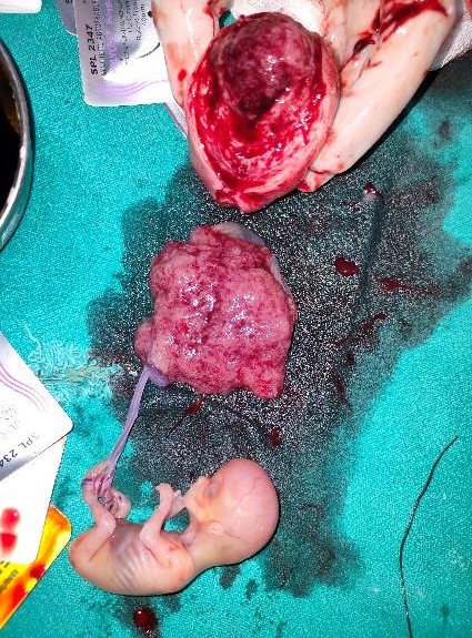
Patient was planned for laparotomy. Intra operative findings were 6*6cms right adnexal mass arising from uterus with evidence of right fallopian tube and right round ligament arising from it-suggestive of right rudimentary horn pregnancy ([Figure 1]) Right uterine horn along with right fallopian tube were removed. No evidence of rupture of the mass. On cut section, evidence of gestational-sac of around 10-12 weeks with evidence of well-formed fetus and placental tissue.([Figure 2]). The rudimentary horn was found to be non communicating.

Whole mass was sent for Histopathological examination. Condition of bilateral ovaries and left fallopian tube were found to be normal. Post operative period was uneventful and patient was discharged on 3rd post-operative day and was advised to follow up after 6 weeks.
Case 2
A 31-year-old female presented at 6 weeks 4 days gestation at Kamla Nehru Hospital for Mother and Child, Indira Gandhi Medical College, Shimla with complain of bleeding and pain in lower abdomen for past 12 days.
The patient was primigravida with history of primary infertility of 7 years for which she had been investigated in MMU, Solan in 2018. Her blood investigations and husband semen analysis were carried out which were found to be normal. Endometrial biopsy was also done which showed no granuloma following which diagnostic laparoscopy proceed chromotubation was performed. It showed free spillage on left side. No comments were given on the right fallopian tube, ovaries and uterus. There was no significant past, medical or surgical history. She had normal menstrual periods with no history of dysmenorrhoea.
At admission, the vitals of the patient were stable with a pulse rate of 98/minute and blood pressure of 106/64mmHg. Her General physical examination was also normal with no pallor and systemic examination was also normal. Her per abdomen examination was also normal with no distension, tenderness, guarding or rigidity. On per speculum examination, minimal bleeding through os was present. On per vaginal examination, uterus was normal in size, soft, non-tender and anteverted, cervical motion tenderness was present, fullness was present in right adnexa, left adnexa was normal, no fullness and nodularity in pouch of Douglas.
Her TVS was carried out in our setup which was a suboptimal study which showed that uterus was normal in size, outline and echotexture with endometrial thickness of 5mm. No gestational sac was seen. Left ovary was normal. Right adnexa showed a cystic structure corresponding to gestational age of 5 weeks 1 day with another cystic structure in it. Right ovary was separately visualised. There was evidence of minimal free fluid in pelvis suggestive of Right adnexal ectopic pregnancy.
Her blood investigations and serum βHCG were sent so as to start her on injection methotrexate considering her to be a candidate for medical management of ectopic pregnancy. Her Hb was 11.6g/dl, blood group was AB +, viral markers were non-reactive and Liver function tests and Renal function tests were within normal limits. Her βHCG was 14,939.5 which was in the higher range for medical management following which she was again subjected to transvaginal ultrasonography.
On repeat TVS, it showed uterus was normal in size, outline and echotexture. No intrauterine pregnancy was seen. Uterus appears to be unicornuate with single cornua in midline. B/L ovaries were normal in size, outline and echotexture. There was an ectopic gestation sac in right adnexa of size 2.6*2.3cm of gestational age 6week 6 days with yolk sac in it. No embryo seen. ? ectopic gestation in right adnexa ? Rudimentary horn ectopic. No free fluid in pouch of douglas.
Following this, patient underwent a laparotomy through a low transverse incision. The operative findings included unruptured ectopic mass of around 2*2 cm in rudimentary horn which was excised and sent for histopathological examination. Uterus, right fallopian tube and bilateral ovaries were grossly normal. Right salpingectomy was done and sent for histopathological examination. There was evidence of thick adhesions between left round ligament and mesentery. Left fallopian tube was tortuous and dye test was negative on left side.


The post-operative period was uneventful and patient was discharged on fourth day. A histological examination confirmed the diagnosis. There was no infiltration of the chorionic villi into the myometrium. Patient was counselled for In Vitro fertilisation.
Case 3
A 30 years old G3P1+1 with 10 weeks 2 days gestation presented in our emergency ward at Kamla Nehru Hospital, Shimla referred from a peripheral rural health center in view of query tubal ectopic pregnancy. Patient came in with chief complains of pain in lower abdomen for past 2 days. The patient had a previous uneventful full term vaginal delivery of a 2.8kg male baby 3 years back at a nearby hospital. The patient had a spontaneous abortion at 6-8 weeks one year later which was medically managed. This was her third pregnancy. Prior to this visit, patient had no antenatal checkup. Patient underwent an ultrasound examination due to pain in lower abdomen at nearby private ultrasound Centre which showed tubal ectopic pregnancy. Patient immediately consulted at peripheral rural health Centre from where patient was referred to our emergency ward. Patient had no remarkable obstetrics or menstrual history and no history of dysmenorrhea. Patient had no significant past, family or medical history.
On admission, the vitals of the patient were stable with blood pressure of 100/66mmHg and pulse of 96 bpm which was regular and good volume. Her general physical examination and systemic examination was also normal with no pallor. On per abdomen examination uterus was soft and just palpable. There was no guarding or rigidity. On per speculum examination a single cervical os seen with os closed and cervical length of around 2.5cms. On per vaginal examination a mass of 12 weeks size felt separately from the uterus with fullness in left adnexa. Right adnexa was normal. No evidence of cervical motion tenderness or fullness in pouch of douglas.
Patient underwent a repeat transvaginal ultrasonography in our institution which showed a normal looking uterus with no gestational sac in it with another gestational sac seen with a viable fetus inside with a crown rump length of 6cms over the left side of uterus with no communication with the main uterine cavity suggestive of left rudimentary horn pregnancy with gestation of 10 weeks.
Patient’s blood investigations were sent. Her hemoglobin was 12gm/dl, her blood group was O positive. Patient underwent a laparotomy through a low transverse incision. The procedure revealed a left rudimentary horn pregnancy. Resection of the rudimentary horn along with ipsilateral fallopian tube was done. A fetus of around 7 cms was found inside. The whole mass was sent for histopathological examination. Post operative period was uneventful and patient was discharged on 4th postoperative day and was advised for follow up.
Discussion
Casper Wolff described the mesonephros in 1759 at the age of 26yrs. The paired structures were named wolffian bodies by the 19th century embryologist, Rathke, in recognition of wolff’s initial discovery and description. The mesonephric (wolffian) and paramesonephric (Mullerian)ducts are discrete primordia that coexist in all embryo upto 8 weeks. The Mullerian ducts develop later into fallopian tubes, uterus and upper portion of vagina in females. Rudimentary horn pregnancy with a unicornuate uterus results due to failure of the complete development of one of the Mullerian ducts and incomplete fusion with the contralateral side.
The ASRM Mullerian anomalies classification 2021(MAC2021)[4] classifies Mullerian anomalies into nine categories which includes Mullerian agenesis, cervical agenesis, unicornuate uterus, uterus didelphys, bircornuate uterus, septate uterus, longitudinal vaginal septum, transverse vaginal septum and complex anomalies. Because Mullerian anomalies represent a continuum of development and many may have combined elements, some anomalies appear in more than one category.
It has been described that rudimentary horn pregnancies are extremely rare, and they are reported at 1:76,000-1:1,60,000 pregnancies. It occurs by transmigration of peritoneal sperms or fertilised ovum in the case of non-communicating uterine horn.[2]
Unicornuate uteri are further subdivided into 2 variants according to the criteria from the American fertility society. Type a includes unicornuate uterus with rudimentary horn and type b unicornuate uterus containing no horn(35%). Type a is further classified into a1 including rudimentary horn containing endometrium and a2 including rudimentary horn with no endometrial cavity (33%). Then a1 is further subdivided into a1a including communicating horn (10%) and a1b a non-communicating horn (22%).[5] In our cases described, all were of the non communicating, cavitary types.
Studies have indicated a vast variation in rupture period, ranging from 5-35 weeks, and that was attributed to the decreased ability of the horn musculature to hypertrophy and dilate as compared to normal uterine musculature. Further it has been identified that around 70-90% of total ruptures occur before 20 weeks and these lead to catastrophic results.[6] As the uterine wall is thicker and more vascular, bleeding is more severe in rudimentary horn pregnancy rupture. A rudimentary horn pregnancy can be further complicated by placenta percreta due to the poorly developed musculature and the small size of the horn; the reported incidence is 11.9%.[7] Fortunately, we were able to diagnose the rudimentary horn pregnancy before rupture.
Most of the times rudimentary horns are asymptomatic. Most cases of rudimentary horn pregnancy provide a diagnostic challenge and are diagnosed after rupture, which leads to emergency surgery, blood transfusions and increased morbidity and mortality.[8]
Diagnosis is difficult in this malformation due to a limited field of ultrasound when compared to other imaging modalities. Sonographic senstivity is only 26% and as the pregnancy advances the specificity goes down.[8] Common misdiagnosis includes tubal pregnancy, cornual pregnancy or abdominal pregnancies as sometimes due to inexpertise of the radiologists, the diagnosis can be missed.
There are three fundamental criteria on ultrasound for the ultimate diagnosis of pregnancy in rudimentary horn. These include pseudopattern of an asymmetrical bicornuate uterus, absent visual continuity between the cervical canal and the lumen of the pregnant horn and the presence of myometrial tissue surrounding the gestational sac.[9]
Some gold standard diagnostic modalities include magnetic resonance imaging (MRI) and 3-D ultrasound in both pregnant and non-pregnant women. An MRI also excludes any other Mullerian or urinary system anomalies in 36% of the cases.[6] We also used 3D ultrasound to confirm our diagnosis in the first case.
Although a definitive treatment is surgical excision of the rudimentary horn so that to prevent rupture and recurrent rudimentary horn pregnancies, other treatment modalities are also being made available. Some authors have described systemic methotrexate administration or feticide with intracardiac potassium chloride as alternatives or adjuncts to surgery in early gestation.[10] Conservative management, until viability is established, has been advocated in selected cases with large myometrial masses. In all such cases, the patient should be informed of the risks of the condition as well as their management options.[11]
Conclusion
Hence, it can be concluded that diagnosing a case of rudimentary horn pregnancy can be a challenge. Most common misdiagnosis can be tubal pregnancy which can delay the necessary intervention required. MRI and diagnostic laparoscopy remain the most effective tools for the diagnosis. Surgical management remains the gold standard which includes excision of the horn prior to rupture with excision of ipsilateral fallopian tube to prevent future ectopic pregnancy.
Source of Funding
None.
Conflict of Interest
None.
References
- S Jomaa, A Ahmad, D Adwan. Successful diagnosis and management of prerupture rudimentary horn pregnancy in the second trimester: a case report. Radiol Case Rep 2021. [Google Scholar]
- K Chatziioannidou, A Fehlmann, J Dubuisson. Case Report: Laparoscopic Management of an Ectopic Pregnancy in a Rudimentary Non-communicating Uterine Horn. Front Surg 2020. [Google Scholar] [Crossref]
- R Jain, N Gami, M Puri, SS Trivedi. A rare case of intact rudimentary horn pregnancy presenting as hemoperitoneum. J Hum Reprod Sci 2010. [Google Scholar]
- S Jacob, L Karunakaran, N Devasia. Rudimentary horn pregnancy: a rare entity. Int J Reprod Contracept Obstet Gynecol 2020. [Google Scholar] [Crossref]
- N Yang, G Fares. Unicornuate uterus. 2022. [Google Scholar] [Crossref]
- DV Kanagal, LC Hanumanalu. Ruptured Rudimentary Horn Pregnancy at 25 Weeks with Previous Vaginal Delivery: A Case Report. Case Rep Obstet Gynecol 2012. [Google Scholar] [Crossref]
- PI Okonta, H Abedi, C Ajuyah, L Omo-Aghoja. Pregnancy in a noncommunicating rudimentary horn of a unicornuate uterus: A case report. Cases J 2009. [Google Scholar] [Crossref]
- DV Kanagal. Ruptured Rudimentary Horn Pregnancy at 25 Weeks with Previous Vaginal Delivery: A Case Report. Case Rep Obstet Gynecol 2012. [Google Scholar] [Crossref]
- S Goyal, A Mahajan, S Goyal. Rudimentary horn of Uterus : A diagnostic challenge in pregnancy. Int J Biomed Adv Res 2017. [Google Scholar]
- SM Pfeifer, M Attaran, J Goldstein, SR Lindheim, JC Petrozza, BW Rackow. ASRM müllerian anomalies classification 2021. Fertil Steril 2021. [Google Scholar]
- E Henriet, H Roman, J Zanati, B Lebreton, JC Sabourin, M Loic. Pregnant noncommunicating rudimentary uterine horn with placenta percreta. JSLS 2008. [Google Scholar]